Epicardial fat, gender, and cardiovascular risk

Submitted: 29 July 2020
Accepted: 4 February 2021
Published: 30 June 2021
Abstract Views: 657
PDF: 340
HTML: 10
Publisher's note
All claims expressed in this article are solely those of the authors and do not necessarily represent those of their affiliated organizations, or those of the publisher, the editors and the reviewers. Any product that may be evaluated in this article or claim that may be made by its manufacturer is not guaranteed or endorsed by the publisher.

Authors

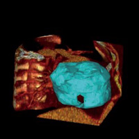
Epicardial fat (EF) is considered an important risk factor and an active player in the pathogenesis of cardiovascular and metabolic diseases. EF is an endocrine organ that releases hormones and mediators, including the circulating C-reactive protein (CRP), and plays a vital role in modifying the vascular endothelial function and promoting the growth of coronary atherosclerosis. This study aimed to investigate the relationship between CRP concentrations and EF in a cohort of patients with metabolic syndrome at risk for coronary artery disease. In our study, carried out in primary prevention, we enrolled 36 subjects (M/F: 21/15; age: 59.3±0.79 yrs) diagnosed with metabolic syndrome. We have classified the patients into two groups: Men and Women. Besides anthropometric characterization and screening laboratory tests, the subjects performed a multidetector computed tomography scan, which allowed the EF quantification. Mean EF was 115.1 cc in the study population. The average EF of women was 111 cc; the average EF of men was 118 cc (P=0.18). CRP levels were strongly positively correlated with EF area in women (P=0.01), while the correlation was not found in men (P=0.4). Our findings suggest that, in women, the EF produces a greater amount of acute-phase proteins and increases the pro-inflammatory state in the epicardial region. For this reason, we can hypothesize, in women, a different role in the development of atherosclerotic plaque of the epicardial fat compared to men.

Dimensions

Altmetric

PlumX Metrics

Downloads

Download data is not yet available.

Citations

Weisberg SP, McCann D, Desai M, et al. Obesity is associated with macrophage accumulation in adipose tissue. J Clin Invest 2003;112:1796-808. DOI: https://doi.org/10.1172/JCI200319246
Xu H, Barnes GT, Yang Q, et al. Chronic inflammation in fat plays a crucial role in the development of obesity-related insulin resistance. J Clin Invest 2003;112:1821-30. DOI: https://doi.org/10.1172/JCI200319451
Mohamed-Ali V, Goodrick S, Rawesh A, et al. Subcutaneous adipose tissue releases interleukin-6, but not tumor necrosis factor-α, in vivo. J Clin Endocrinol Metab 1997;82:4196-200. DOI: https://doi.org/10.1210/jc.82.12.4196
Corradi D, Maestri R, Callegari S, et al. The ventricular epicardial fat is related to the myocardial mass in normal, ischemic and hypertrophic hearts. Cardiovasc Pathol 2004;13:313-6. DOI: https://doi.org/10.1016/j.carpath.2004.08.005
Iacobellis G, Malavazos AE, Corsi MM. Epicardial fat: from the biomolecular aspects to the clinical practice. Int J Biochem Cell Biol 2011;43:1651-4.
Chen X, Jiao Z, Wang L, et al. Roles of human epicardial adipose tissue in coronary artery atherosclerosis. J Huazhong Univ Sci Technolog Med Sci 2010;30:589-93. DOI: https://doi.org/10.1007/s11596-010-0547-9
Monti M, Monti A, Murdolo G, et al. Correlation between epicardial fat and cigarette smoking: CT imaging in patients with metabolic syndrome. Scand Cardiovasc J 2014;48:317-22. DOI: https://doi.org/10.3109/14017431.2014.942872
Al-Delaimy WK, Willett WC, Manson JE, et al. Smoking and mortality among women with type 2 diabetes: the Nurses’ Health Study cohort. Diabetes Care 2001;24:2043-8. DOI: https://doi.org/10.2337/diacare.24.12.2043
Dey D, Nakazato R, Li D, Berman SD. Epicardial and thoracic fat - Noninvasive measurement and clinical implications. Cardiovasc Diagn Ther 2012;2:85-93.
Flüchter S, Haghi D, Dinter D, et al. W Volumetric assessment of epicardial adipose tissue with cardiovascular magnetic resonance imaging. Obesity (Silver Spring) 2007;15:870-8. DOI: https://doi.org/10.1038/oby.2007.591
Mosteller RD. Simplified calculation of body-surface area. N Engl J Med 1987;317:1098. DOI: https://doi.org/10.1056/NEJM198710223171717
Friedewald WT, Levy RI, Fredrickson DS. Estimation of the concentration of low-density lipoprotein cholesterol in plasma, without use of the preparative ultracentrifuge. Clin Chem 1972;18,499-502. DOI: https://doi.org/10.1093/clinchem/18.6.499
Bland JM, Altman DG Statistical methods for assessing agreement between two methods of clinical measurement. Lancet 1986;8:307-10. DOI: https://doi.org/10.1016/S0140-6736(86)90837-8
Shioji K, Moriguchi A, Moriwaki S, et al. Hypoadiponectinemia implies the development of atherosclerosis in carotid and coronary arteries. J Cardiol 2005;46:105-12.
Libby P, Ridker PM, Maseri A. Inflammation and atherosclerosis. Circulation 2002;105:1135-43. DOI: https://doi.org/10.1161/hc0902.104353
Greenstein AS, Khavandi K, Withers SB, et al. Local inflammation and hypoxia abolish the protective anticontractile properties of perivascular fat in obese patients. Circulation 2009;119:1661-70. DOI: https://doi.org/10.1161/CIRCULATIONAHA.108.821181
Greif M, Becker A, von Ziegler F, et al. Pericardial adipose tissue determined by dual source CT is a risk factor for coronary atherosclerosis. Arterioscler Thromb Vasc Biol 2009;29:781-6. DOI: https://doi.org/10.1161/ATVBAHA.108.180653
Salgado-Somoza A, Teijeira-Fernández E, Rubio J, et al. Coronary artery disease is associated with higher epicardial retinol-binding protein 4 (RBP4) and lower glucose transporter (GLUT) 4 levels in epicardial and subcutaneous adipose tissue. Clin Endocrinol (Oxf) 2012;76:51-8. DOI: https://doi.org/10.1111/j.1365-2265.2011.04140.x
Ridker PM, Buring JE, Shih J, et al. Prospective study of C-reactive protein and the risk of future cardiovascular events among apparently healthy women. Circulation 1998;98:731-3. DOI: https://doi.org/10.1161/01.CIR.98.8.731
Hartman J, Frishman WH. Inflammation and atherosclerosis: a review of the role of interleukin-6 in the development of atherosclerosis and the potential for targeted drug therapy. Cardiol Rev 2014;22:147-51. DOI: https://doi.org/10.1097/CRD.0000000000000021
Burke AP, Tracy RP, Kolodgie F et al. Elevated C-reactive protein values and atherosclerosis in sudden coronary death: association with different pathologies. Circulation 2002;105:2019-23. DOI: https://doi.org/10.1161/01.CIR.0000015507.29953.38
Pasceri V, Chang J, Willerson JT et al. Modulation of C-reactive protein-mediated monocyte chemoattractant protein-1 induction in human endothelial cells by anti-atherosclerosis drugs. Circulation 2001;103:2531-4. DOI: https://doi.org/10.1161/01.CIR.103.21.2531
Després JP, Pascot A, Bergeron J, et al. Abdominal obesity: the critical correlate of elevated plasma C-reactive protein levels associated with features of the insulin resistance syndrome in men. Atherosclerosis 2000;151:98. DOI: https://doi.org/10.1016/S0021-9150(00)80443-8
Saijo Y, Kiyota N, Kawasaki Y, et al. Relationship between C reactive protein and visceral adipose tissue in healthy Japanese subjects. Diabetes Obes Metab 2004;6:249-58. DOI: https://doi.org/10.1111/j.1462-8902.2003.0342.x
Danesh J, Collins R, Appleby P, et al. Association of fibrinogen, C-reactive protein, albumin, or leukocyte count with coronary heart disease: meta-analyses of prospective studies. JAMA 1998;279:1477-82. DOI: https://doi.org/10.1001/jama.279.18.1477
Pérez-Segura P, de Dios O, Herrero L, et al. Children with type 1 diabetes have elevated high-sensitivity C-reactive protein compared with a control group. BMJ Open Diabetes Res Care 2020;8:e001424. DOI: https://doi.org/10.1136/bmjdrc-2020-001424
Miyazawa I, Ohkubo T, Kadowaki S, et al. Change in pericardial fat volume and cardiovascular risk factors in a general population of Japanese men. Circ J 2018;82:2542-8. DOI: https://doi.org/10.1253/circj.CJ-18-0153
Hak AE, Stehouwer CD, Bots ML, et al. Associations of C-reactive protein with measures of obesity, insulin resistance, and subclinical atherosclerosis in healthy, middle-aged women. Arterioscler Thromb Vasc Biol 1999;19:1986-91. DOI: https://doi.org/10.1161/01.ATV.19.8.1986
Ray P, Ghosh SK, Zhang DH, et al. Repression of interleukin-6 gene expression by 17 inhibition of the DNA-binding activity of the transcription NF-IL6 and NF-kappa B by the estrogen receptor. FEBS Lett 1997;409:79-85. DOI: https://doi.org/10.1016/S0014-5793(97)00487-0
Van Baal WM, Kenemans P, van der Mooren MJ, et al. Increased C-reactive protein levels during short-term hormone replacement therapy in healthy postmenopausal women. Thromb Haemost 1999;81:925-8. DOI: https://doi.org/10.1055/s-0037-1614600
White UA, Tchoukalova YD. Sex dimorphism and depot differences in adipose tissue function. Biochim Biophys Acta 2014;1842:377-92. DOI: https://doi.org/10.1016/j.bbadis.2013.05.006
Iacobellis G, Malavazos AE, Corsi MM. Epicardial fat: from the biomolecular aspects to the clinical practice. Int J Biochem Cell Biol 2011;43:1651-4. DOI: https://doi.org/10.1016/j.biocel.2011.09.006
Gruzdeva O, Uchasova E, Dyleva Y, et al. Adipocytes directly affect coronary artery disease pathogenesis via induction of adipokine and cytokine imbalances. Front Immunol 2019;10:2163. DOI: https://doi.org/10.3389/fimmu.2019.02163

How to Cite

Monti, M., Marchetti, R., & Vincentelli, G. M. (2021). Epicardial fat, gender, and cardiovascular risk. Italian Journal of Medicine, 15(2). https://doi.org/10.4081/itjm.2021.1349

Similar Articles

You may also start an advanced similarity search for this article.