Smad2/3 signaling involved in urotensin II-induced phenotypic differentiation, collagen synthesis and migration of rat aortic adventitial fibroblasts

Published: 30 August 2023
Abstract Views: 487
PDF: 203
HTML: 18
Publisher's note
All claims expressed in this article are solely those of the authors and do not necessarily represent those of their affiliated organizations, or those of the publisher, the editors and the reviewers. Any product that may be evaluated in this article or claim that may be made by its manufacturer is not guaranteed or endorsed by the publisher.

Authors

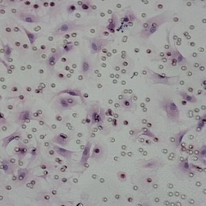
Objective. To investigate whether Smad2/3 signaling is involved in urotensin II (UII) induced activation of aortic adventitial fibroblasts. Materials and Methods. Growth-arrested adventitial fibroblasts were stimulated with UII in the presence or absence of urotensin II receptor (UT) antagonist SB710411 or transfected with Smad2/3 small inhibitory RNA (siRNA). UII stimulated Smad2/3 phosphorylation, α-smooth muscle actin (α-SMA), and collagen I expression and migration of adventitial fibroblasts were evaluated by western blot analysis, real-time reverse transcription polymerase chain reaction, immunofluorescence, ELISA, and transwell migration assay, respectively. Results. In cultured adventitial fibroblasts, UII time- and dose-dependently stimulated Smad2/3 protein phosphorylation, with maximal effect at 10-8 mol/l (increased by 147.2%, P<0.001). UII stimulated Smad2/3 upregulation and nuclear translocation. SB710411 significantly inhibited these effects. In addition, UII potently induced α-SMA and procollagen 1 protein or mRNA expression (P<0.01), which were completely blocked by Smad2 (decreased by 75.1%, 54.2% in protein, and by 73.3% and 38.2% in mRNA, respectively, P<0.01) or Smad3 siRNA (decreased by 80.3% and 47.0% in protein, and by 72.3% and 47.7% in mRNA, respectively, P<0.01). Meanwhile, Smad2 or smad3 siRNA significantly inhibited the UII-induced collagen 1 secretion and cell migration. Conclusions. UII may stimulate adventitial-fibroblast phenotype conversion, migration, and collagen I synthesis via phosphorylated-Smad2/3 signal transduction pathways.

Dimensions

Altmetric

PlumX Metrics

Downloads

Download data is not yet available.

Citations

Rodriguez-Vita J, Sanchez-Lopez E, Esteban V, et al. Angiotensin II activates the Smad pathway in vascular smooth muscle cells by a transforming growth factorbeta-independent mechanism. Circulation 2005;111:2509-17. DOI: https://doi.org/10.1161/01.CIR.0000165133.84978.E2
Liu Y, Liang C, Liu X, et al. AGEs increased migration and inflammatory responses of adventitial fibroblasts via RAGE, MAPK and NF-kappaB pathways. Atherosclerosis 2010;208:34-42. DOI: https://doi.org/10.1016/j.atherosclerosis.2009.06.007
Lan TH, Huang XQ, Tan HM. Vascular fibrosis in atherosclerosis. Cardiovasc Pathol 2013;22:401-7. DOI: https://doi.org/10.1016/j.carpath.2013.01.003
Stenmark KR, Yeager ME, El KK, et al. The adventitia: essential regulator of vascular wall structure and function. Annu Rev Physiol 2013;75:23-47. DOI: https://doi.org/10.1146/annurev-physiol-030212-183802
Wu X, Lu Q. Expression and significance of alpha-SMA and PCNA in the vascular adventitia of balloon-injured rat aorta. Exp Ther Med 2013;5:1671-6. DOI: https://doi.org/10.3892/etm.2013.1059
Evans RA, Tian YC, Steadman R, Phillips AO. TGFbeta1-mediated fibroblast-myofibroblast terminal differentiation-the role of Smad proteins. Exp Cell Res 2003;282:90-100. DOI: https://doi.org/10.1016/S0014-4827(02)00015-0
Forte A, Della CA, De Feo M, et al. Role of myofibroblasts in vascular remodelling: focus on restenosis and aneurysm. Cardiovasc Res 2010;88:395-405. DOI: https://doi.org/10.1093/cvr/cvq224
Boyd R, Ratsep MT, Ding LL, Wang HD. ETA and ETB receptors are expressed in vascular adventitial fibroblasts. Am J Physiol Heart Circ Physiol 2011;301:H2271-H2278. DOI: https://doi.org/10.1152/ajpheart.00869.2010
Song N, Ding W, Chu S, et al. Urotensin II stimulates vascular endothelial growth factor secretion from adventitial fibroblasts in synergy with angiotensin II. Circ J 2012;76:1267-73. DOI: https://doi.org/10.1253/circj.CJ-11-0870
Zhang YG, Hu YC, Mao YY, et al. Transforming growth factor-beta1 involved in urotensin II-induced phenotypic differentiation of adventitial fibroblasts from rat aorta. Chin Med J (Engl) 2010;123:3634-9.
Di Wang H, Ratsep MT, Chapman A, Boyd R. Adventitial fibroblasts in vascular structure and function. the role of oxidative stress and beyond. Can J Physiol Pharmacol 2010;88:177-86. DOI: https://doi.org/10.1139/Y10-015
Zhang YG, Li YG, Wei RH, et al. Urotensin II is an autocrine/paracrine growth factor for aortic adventitia of rat. Regul Pept 2008;151:88-94. DOI: https://doi.org/10.1016/j.regpep.2008.10.004
Shi L, Ding W, Li D, et al. Proliferation and anti-apoptotic effects of human urotensin II on human endothelial cells. Atherosclerosis 2006;188:260-4. DOI: https://doi.org/10.1016/j.atherosclerosis.2005.10.044
Vaudry H, Leprince J, Chatenet D, et al. International Union of Basic and Clinical Pharmacology. XCII. Urotensin II, urotensin II-related peptide, and their receptor: from structure to function. Pharmacol Rev 2015;67:214-58. DOI: https://doi.org/10.1124/pr.114.009480
Zhang YG, Li J, Li YG, Wei RH. Urotensin II induces phenotypic differentiation, migration, and collagen synthesis of adventitial fibroblasts from rat aorta. J Hypertens 2008;26:1119-26. DOI: https://doi.org/10.1097/HJH.0b013e3282fa1412
Shiraishi Y, Watanabe T, Suguro T, et al. Chronic urotensin II infusion enhances macrophage foam cell formation and atherosclerosis in apolipoprotein Eknockout mice. J Hypertens 2008;26:1955-65. DOI: https://doi.org/10.1097/HJH.0b013e32830b61d8
Zhao J, Ding W, Song N, et al. Urotensin II-induced collagen synthesis in cultured smooth muscle cells from rat aortic media and a possible involvement of transforming growth factor-beta1/Smad2/3 signaling pathway. Regul Pept 2013;182:53-8. DOI: https://doi.org/10.1016/j.regpep.2012.12.006
Tzanidis A, Hannan RD, Thomas WG, et al. Direct actions of urotensin II on the heart: implications for cardiac fibrosis and hypertrophy. Circ Res 2003;93:246-53. DOI: https://doi.org/10.1161/01.RES.0000084382.64418.BC
Wang W, Huang XR, Canlas E, et al. Essential role of Smad3 in angiotensin II-induced vascular fibrosis. Circ Res 2006;98:1032-9. DOI: https://doi.org/10.1161/01.RES.0000218782.52610.dc
Rodrigues DR, Rodrigues-Diez R, Lavoz C, et al. Statins inhibit angiotensin II/Smad pathway and related vascular fibrosis, by a TGF-beta-independent process. PLoS One 2010;5:e14145. DOI: https://doi.org/10.1371/journal.pone.0014145
Javelaud D, Mauviel A. Mammalian transforming growth factor-betas: Smad signaling and physio-pathological roles. Int J Biochem Cell Biol 2004;36:1161-5. DOI: https://doi.org/10.1016/S1357-2725(03)00255-3
Yang F, Chung AC, Huang XR, Lan HY. Angiotensin II induces connective tissue growth factor and collagen I expression via transforming growth factor-beta-dependent and -independent Smad pathways: the role of Smad3. Hypertension 2009;54:877-84. DOI: https://doi.org/10.1161/HYPERTENSIONAHA.109.136531
Li JH, Huang XR, Zhu HJ, et al. Advanced glycation end products activate Smad signaling via TGF-betadependent and independent mechanisms: implications for diabetic renal and vascular disease. FASEB J 2004;18:176-8. DOI: https://doi.org/10.1096/fj.02-1117fje
Dai HY, Kang WQ, Wang X, et al. The involvement of transforming growth factor-beta1 secretion in urotensin II-induced collagen synthesis in neonatal cardiac fibroblasts. Regul Pept 2007;140:88-93. DOI: https://doi.org/10.1016/j.regpep.2006.11.015
Tran L, Kompa AR, Kemp W, et al. Chronic urotensin-II infusion induces diastolic dysfunction and enhances collagen production in rats. Am J Physiol Heart Circ Physiol 2010;298:H608-H613. DOI: https://doi.org/10.1152/ajpheart.00942.2009
Matsusaka S, Wakabayashi I. Enhancement of vascular smooth muscle cell migration by urotensin II. Naunyn Schmiedebergs Arch Pharmacol 2006;373:381-6. DOI: https://doi.org/10.1007/s00210-006-0086-x
Xu S, Jiang H, Wu B, et al. Urotensin II induces migration of endothelial progenitor cells via activation of the RhoA/Rho kinase pathway. Tohoku J Exp Med 2009;219:283-388. DOI: https://doi.org/10.1620/tjem.219.283
Zhang YG, Kuang ZJ, Mao YY, et al. Osteopontin is involved in urotensin II-induced migration of rat aortic adventitial fibroblasts. Peptides 2011;32:2452-8. DOI: https://doi.org/10.1016/j.peptides.2011.10.018
Hinz B. Formation and function of the myofibroblast during tissue repair. J Invest Dermatol 2007;127:526-37. DOI: https://doi.org/10.1038/sj.jid.5700613
Desmouliere A, Darby IA, Gabbiani G. Normal and pathologic soft tissue remodeling. role of the myofibroblast, with special emphasis on liver and kidney fibrosis. Lab Invest 2003;83:1689-707. DOI: https://doi.org/10.1097/01.LAB.0000101911.53973.90

Supporting Agencies

National Natural Science Foundation of China, Natural Science Foundation of Guangdong Province of China

How to Cite

Lin, W., Yang, X., Zheng, F., Yang, J., & Zhang, Y. (2023). Smad2/3 signaling involved in urotensin II-induced phenotypic differentiation, collagen synthesis and migration of rat aortic adventitial fibroblasts. Italian Journal of Medicine, 17(2). https://doi.org/10.4081/itjm.2023.1637